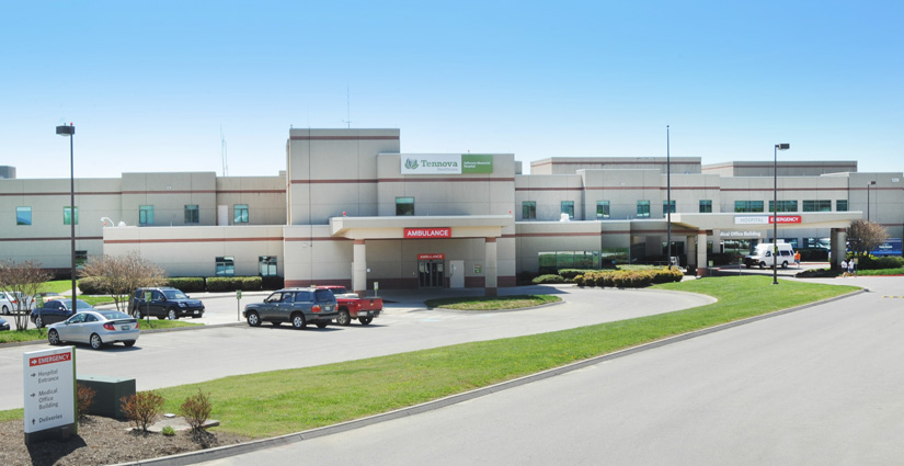
Patient Price Information List
Disclaimer: Jefferson Memorial Hospital determines its standard charges for patient items and services through the use of a chargemaster system, which is a list of charges for the components of patient care that go into every patient’s bill. These are the baseline rates for items and services provided at the Hospital. The chargemaster is similar in concept to the manufacturer’s suggested retail price (“MSRP”) on a particular product or good. The charges listed provide only a general starting point in determining the potential costs of an individual patient’s care at the Hospital. This list does not reflect the actual out-of-pocket costs that may be paid by a patient for any particular service, it is not binding, and the actual charges for items and services may vary.
Many factors may influence the actual cost of an item or service, including insurance coverage, rates negotiated with payors, and so on. Government payors, such as Medicare and Medicaid for example, do not pay the chargemaster rates, but rather have their own set rates that hospitals are obligated to accept. Commercial insurance payments are based on contract negotiations with payors and may or may not reflect the standard charges. The cost of treatment also may be impacted by variables involved in a patient’s actual care, such as specific equipment or supplies required, the length of time spent in surgery or recovery, additional tests, or any changes in care or unexpected conditions or complications that arise. Moreover, the foregoing list of charges for services only includes charges from the Hospital. It does not reflect the charges for physicians, such as the surgeon, anesthesiologist, radiologist, pathologist, or other physician specialists or providers who may be involved in providing particular services to a patient. These charges are billed separately.
Individuals with questions about their out-of-pocket costs of service and other financial information should contact the hospital or consider contacting their insurers for further information.
Jefferson Memorial Hospital Patient Information Price List
LOCAL MARKET HOSPITALS
In order to present a meaningful comparison, Jefferson Memorial Hospital has partnered with Hospital Pricing Specialists LLC to analyze current charges, based off CMS adjudicated claims through 3/31/18. Jefferson Memorial Hospital charges are displayed and compared with the local market charge, consisting of the following hospitals:
Fort Loudon Medical Center
Lenoir City
TN
Laughlin Memorial Hospital
Greeneville
TN
Roane Medical Center
Harriman
TN
Takoma Regional Hospital
Greeneville
TN
Tennova La Follette Medical Ctr
La Follette
TN
Jefferson Memorial Hospital Patient Information Price List
INPATIENT ROOM AND BOARD DAILY CHARGES
Description
Variance
Private Room
Private Room
27% higher than market
Intensive Care Unit
Intensive Care Unit
4% higher than market
Jefferson Memorial Hospital Patient Information Price List
OUTPATIENT EMERGENCY DEPARTMENT CHARGES
Emergency Department charges are based on the level of emergency care provided to our patients. The levels, with Level 1 representing basic emergency care, reflect the type of accommodations needed, the personnel resources, the intensity of care and the amount of time needed to provide treatment. The following charges do not include fees for drugs, supplies or additional ancillary procedures that may be required for a particular emergency treatment. They also do not include fees for Emergency Department physicians, who will bill separately for their services.
Description
Variance
Emergency Critical Care, Each Additional 30 Minutes
Critical care, evaluation and management of the critically ill or critically injured patient; each additional 30 minutes
66% lower than market
Emergency Department Visit - Level 1
Emergency department visit for the evaluation and management of a patient, which requires these 3 key components: A problem focused history; A problem focused examination; and Straightforward medical decision making. Counseling and/or coordination of care with other physicians, other qualified health care professionals, or agencies are provided consistent with the nature of the problem(s) and the patient's and/or family's needs. Usually, the presenting problem(s) are self limited or minor.
27% higher than market
Emergency Department Visit - Level 2
Emergency department visit for the evaluation and management of a patient, which requires these 3 key components: An expanded problem focused history; An expanded problem focused examination; and Medical decision making of low complexity. Counseling and/or coordination of care with other physicians, other qualified health care professionals, or agencies are provided consistent with the nature of the problem(s) and the patient's and/or family's needs. Usually, the presenting problem(s) are of low to moderate severity.
43% higher than market
Emergency Department Visit - Level 3
Emergency department visit for the evaluation and management of a patient, which requires these 3 key components: An expanded problem focused history; An expanded problem focused examination; and Medical decision making of moderate complexity. Counseling and/or coordination of care with other physicians, other qualified health care professionals, or agencies are provided consistent with the nature of the problem(s) and the patient's and/or family's needs. Usually, the presenting problem(s) are of moderate severity.
38% higher than market
Emergency Department Visit - Level 4
Emergency department visit for the evaluation and management of a patient, which requires these 3 key components: A detailed history; A detailed examination; and Medical decision making of moderate complexity. Counseling and/or coordination of care with other physicians, other qualified health care professionals, or agencies are provided consistent with the nature of the problem(s) and the patient's and/or family's needs. Usually, the presenting problem(s) are of high severity, and require urgent evaluation by the physician or other qualified health care professionals but do not pose an immediate significant threat to life or physiologic function.
46% higher than market
Emergency Department Visit - Level 5
Emergency department visit for the evaluation and management of a patient, which requires these 3 key components within the constraints imposed by the urgency of the patient's clinical condition and/or mental status: A comprehensive history; A comprehensive examination; and Medical decision making of high complexity. Counseling and/or coordination of care with other physicians, other qualified health care professionals, or agencies are provided consistent with the nature of the problem(s) and the patient's and/or family's needs. Usually, the presenting problem(s) are of high severity and pose an immediate significant threat to life or physiologic function.
43% higher than market
Jefferson Memorial Hospital Patient Information Price List
OUTPATIENT OCCUPATIONAL THERAPY CHARGES
The following charges reflect the most common services offered by our Occupational Therapy department. Patients may have additional charges, depending on the services performed.
Description
Variance
Paraffin Wax Bath
Paraffin Wax Bath
A paraffin bath may be used on small, irregular surfaces such as the wrists, hands, and feet to treat acute or chronic pain and stiffness. Paraffin is a mineral wax derived from petroleum with a low melting point, which allows extended contact with the skin without the risk of thermal injury. Paraffin is melted in a small tub and the extremity is immersed in the liquid. The moist heat from the wax increases blood flow to the area and relaxes muscle tissue. Paraffin bath treatments may be used prior to exercise to reduce joint stiffness and increase range of motion. This treatment modality is helpful for patients with osteoarthritis, rheumatoid conditions, fibromyalgia, and scleroderma. Paraffin baths may also be prescribed for bursitis, tendonitis, and muscle sprains or strains.
5% higher than market
Jefferson Memorial Hospital Patient Information Price List
OUTPATIENT PULMONARY THERAPY CHARGES
The following charges reflect the most common services offered by our Pulmonary Therapy department. Patients may have additional charges, depending on the services performed.
Description
Variance
CPAP Initiation & Management
Continuous positive airway pressure (CPAP) ventilation is used primarily to treat sleep apnea. It may also be prescribed to treat preterm infants whose lungs have not fully developed. CPAP uses a mask or other breathing device that fits over the nose and mouth which is connected via a tube to a CPAP device. The CPAP machine delivers an air mixture at a continuous low level of pressure. The continuous positive airway pressure keeps the airways open and prevents mechanical obstruction of the flow of air caused by relaxation and collapse of airway structures during sleep. This code is used for initial set-up and management. A durable medical device provider delivers the CPAP device and other required equipment to the home or a residential facility. The device is set up and programmed to the settings indicated by the written prescription obtained from the physician or other qualified health care professional. The patient or caregiver is instructed on correct use of the CPAP and then is asked to demonstrate understanding by placing the mask over the mouth and nose and turning on the machine.
3% lower than market
Holter Testing - 48-Hour EKG
Electrocardiographic (ECG) rhythm-derived data is gathered for up to 48 hours of monitoring as the patient goes about regular daily activity while wearing an external ECG recording device, also called a Holter monitor. Electrodes or leads are placed on the patient's chest, and the patient is instructed on the use of the monitor. The recording device makes continuous, original ECG wave recordings for a 12 to 48 hour period. The recordings are captured on magnetic tape or digitized medium to be reviewed later. At the end of the recording period, the patient returns to the office with the device. Stored data derived from the continuous recordings of the electrical activity of the heart include heart rhythm and rate, ST analysis, variability in heart rate and T-wave alternans. Visual superimposition scanning is done to give a 'page review' of the entire recording, identifying different ECG waveforms with selective samples of rhythm strips. A report is made after analysis of the scanning, and the physician or other qualified health care professional reviews and interprets the data for heart arrhythmias.
19% lower than market
Jefferson Memorial Hospital Patient Information Price List
OUTPATIENT LABORATORY AND PATHOLOGY CHARGES
The following charges reflect our most common laboratory procedures. For all lab specimens collected via blood draw, the venipuncture will be charged separately.
Description
Variance
Albumin Level
Albumin Level
A blood test is performed to measure albumin levels in serum, plasma, or whole blood. Albumin is a plasma protein responsible for regulating the colloidal osmotic pressure of blood. It is capable of binding water, electrolytes (sodium, potassium, calcium), fatty acids, hormones, bilirubin, and drugs/medications. Albumin levels are used to assess nutritional status. A blood sample is obtained by separately reportable venipuncture. The plasma, serum or whole blood is tested using spectrophotometry or quantitative nephelometry.
20% higher than market
Alpha-Fetoprotein (AFP) Level, Serum
Alpha-fetoprotein (AFP) is measured in serum or amniotic fluid during pregnancy to screen for neural tube defects such as spina bifida and anencephaly, chromosomal abnormalities such as Down syndrome or Edwards syndrome, and omphalocele. AFP is a protein produced by the yolk sac of the fetus in early gestation and then later by the liver and gastrointestinal tract of the fetus. A blood sample is obtained by separately reportable venipuncture between 14 and 22 weeks gestation. The blood (serum) test screens for high and low levels of AFP. High levels are associated with neural tube defects while low levels are associated with chromosomal abnormalities.
4% lower than market
Amikacin (antibiotic) level
2% lower than market
Ammonia Level
Ammonia Level
A blood test is performed to measure ammonia levels. Ammonia is a by product of protein metabolism and is normally converted to urea by the liver and excreted via the kidney. Elevated ammonia levels may result from cirrhosis or hepatitis. Symptoms of elevated ammonia levels are confusion, tremors, excessive sleepiness, or coma. Testing may be performed in disease states such as Reyes syndrome or liver failure. A blood sample is obtained by a separately reportable venipuncture or arterial access line. The specimen is then tested using colorimetry.
10% higher than market
Analysis for Gastrointestinal Bacteria
A laboratory test is performed to measure Helicobacter pylori antibodies. H. pylori is a bacterium found in contaminated food and water and may be spread person to person. The organism causes chronic inflammation of the gastrointestinal mucosa, primarily in the stomach and duodenum, which leads to erosion or ulcer formation. The seroconversion of IgA and IgG antibodies will occur together within about 60 days of exposure. Elevated titers to both IgA and IgG antibodies in the presence of symptoms may represent an active H. pylori infection. The antibody test should be confirmed with bacterial isolation or other diagnostic tests such as H. pylori breath test, or fecal antigen by EIA. It is also possible to test for IgM antibodies, however the clinical usefulness of this measurement has not been clearly established. A blood sample is obtained by separately reportable venipuncture. Serum is tested for IgA/IgG antibodies using semi-quantitative enzyme immunoassay and for IgM antibodies using semi-quantitative enzyme-linked immunosorbent assay.
24% lower than market
Analysis for antibody bacteria
8% higher than market
Analysis for antibody to Diphtheria (bacteria)
20% higher than market
Analysis for antibody to HIV-1 and HIV-2 virus
Lab test for HIV-1/HIV-2
41% lower than market
Analysis for antibody to tetanus bacteria (Clostridium tetanus)
19% higher than market
Analysis of Substance Using Immunoassay Technique
An immunoassay is performed to detect a chemical substance (analyte) other than an antigen or antibody produced by the body in response to an infectious agent. There are dozens of immunoassays for non-infectious antigens and antibodies that are reported with these codes. Examples of non-infectious diseases and disorders that may be worked up using these tests include: celiac disease, motor and sensory neuropathy, Crohn's disease or other inflammatory bowel disease, liver disease, thyroid disease. Examples of specific antigens and antibodies reported with these codes include: Asialo IgG and IgM, ganglioside IgG and IgM, gliadin IgA and IgG. Codes are reported for each immunoglobulin class or subclass tested using this technique, so for example when testing for gliadin IgA and IgG by immunoassay, the code would be reported twice. Qualitative or semiquantitative testing is performed. Qualitative tests detect only the presence or absence of the specific substance. Semiquantitative tests provide a rough estimate of the amount of the substance being tested.
15% lower than market
Antithrombin III antigen (clotting inhibitor) level
14% lower than market
Apolipoprotein level
24% lower than market
Automated white blood cell count
41% lower than market
Bacterial Culture, Any Source Blood
A tissue sample is collected and placed in a medium conducive to the growth of bacteria. The culture is examined for the growth of bacteria that can survive without oxygen.
39% lower than market
Bacterial urine culture; quantitative colony count
"A laboratory test is performed to determine the presence or absence of bacterial colonies in urine and provide a colony count. Bacteria in urine may indicate an acute or chronic urinary tract infection (UTI) including pyelonephritis, cystitis, urethritis, or acute urethral syndrome. A urine sample is obtained by clean catch, mid-stream void or catheterization. Using a calibrated loop, the urine specimen is inoculated onto agar plates and incubated. Quantitative colony counts are determined and potential pathogens are identified. A colony count 10,000 cfu/mL is reported as ""organism present"" and may indicate an infection. Comingled flora of the urethra and mixed organisms in the colony counts are reported as ""mixed flora"" and most often represent contamination."
19% lower than market
Bacterial culture for anaerobic isolates
17% higher than market
Bilirubin Level; Total
A laboratory test is performed to measure total or direct bilirubin. Bilirubin is a pigmented waste product normally produced when red blood cells (RBCs) break down. Non-water soluble (unconjugated) bilirubin is carried on albumin to the liver where it attaches to sugar molecules to become conjugated. Conjugated (direct) bilirubin is water soluble and able to pass from the liver to the small intestine. Further breakdown of bilirubin occurs in the small intestine and it is eventually eliminated in the feces in the form of stercobilin. Total bilirubin is the sum of conjugated (direct) and unconjugated bilirubin. A test for either conjugated or direct bilirubin or total bilirubin may be ordered to diagnose and monitor liver disorders, hemolytic anemia, and newborn (physiologic) jaundice. A blood sample is obtained by separately reportable venipuncture or heel stick. Other body fluids, including cerebral spinal fluid, may be collected and tested for total bilirubin. Serum/plasma is tested using quantitative spectrophotometry.
6% lower than market
Blood Creatinine
Blood Creatinine
A blood sample is taken to measure creatinine levels. Creatinine is a waste product produced by the muscles in the breakdown of creatine, which is a compound used by the muscles to create energy for contraction. The waste product, creatinine, is excreted by the kidneys and blood levels provide a good measurement of renal function. Creatinine may be checked to screen for or monitor treatment of renal disease. Creatinine levels may also be monitored in patients with acute or chronic illnesses that may impair renal function and in patients on medications that affect renal function. Creatinine is measured using spectrophotometry.
5% higher than market
Blood Glucose Level Test
A blood sample is obtained to measure total (quantitative) blood glucose level. Glucose is a simple sugar that is the main source of energy for the body. Carbohydrates are broken down into simple sugars, primarily glucose, absorbed by the intestine, and circulated in the blood. Insulin, a hormone produced by the pancreas, regulates glucose level in the blood and transports glucose to cells in other tissues and organs. When more glucose is available in the blood than is required, it is converted to glycogen and stored in the liver or converted to fat and stored in adipose (fat) tissue. If the glucose/insulin metabolic process is working properly, blood glucose will remain at a fairly constant, healthy level. Glucose is measured to determine whether the glucose/insulin metabolic process is functioning properly. It is used to monitor glucose levels and determine whether they are too low (hypoglycemia) or too high (hyperglycemia) as well as test for diabetes and monitor blood sugar control in diabetics.
13% higher than market
Blood Potassium Level
A blood sample is obtained to measure potassium level. Potassium is a positively charged electrolyte that works in conjunction with other electrolytes, such as sodium, chloride, and carbon dioxide (CO2), to regulate body fluid, stimulate muscle contraction, and maintain proper acid-base balance. Potassium is found in all body fluids but mostly stored within cells, not in extracellular fluids, blood serum, or plasma. Small fluctuations in blood potassium, either too high (hyperkalemia) or too low (hypokalemia), can have serious, even life-threatening, consequences. Potassium level is used to screen for and monitor renal disease; monitor patients on certain medications, such as diuretics, as well as patients with acute and chronic conditions, such as dehydration or endocrine disorders. Because blood potassium affects heart rhythm and respiratory rate, it is routinely checked prior to major surgical procedures. Potassium is measured by ion-selective electrode (ISE) methodology.
8% higher than market
Calcitonin (hormone) level
16% higher than market
Calcium Level
Calcium Level
A blood sample is taken to measure the amount of total calcium. Calcium is one of the most important minerals in the body. About 99 percent of the calcium found the body is stored in the bones. The remaining one percent circulates in the blood. Calcium may be ionized (free) or attached (bound) to proteins. Free calcium is the calcium metabolically active in the body. Bound calcium is inactive. Total calcium is a measurement of the total amount of both free calcium and bound calcium circulating in the blood. Total calcium is measured to screen for or monitor a number of conditions, including those affecting the bones, heart, nerves, kidneys and teeth. Total calcium is measured using spectrophotometry.
13% higher than market
Carbamazepine Level
Carbamazepine Level
A laboratory test is performed to determine total or free carbamazepine levels. Carbamazepine, also referred to as Tegretol, is an anticonvulsant used to treat epilepsy and may also be used as an analgesic to treat trigeminal neuralgia. Carbamazepine, carbamazepine metabolite (10,11-epoxide), and free carbamazepine are routinely measured to determine optimal doses in patients with epilepsy as well as to monitor for carbamazepine toxicity. A blood sample is obtained by separately reportable venipuncture. Blood serum is then tested using one of several techniques including high performance liquid chromatography or fluorescent polarization immunoassay. Total carbamazepine tests the total amount present in the blood. Under normal circumstances, circulating carbamazepine is 75% protein bound. In some patients carbamazepine may be displaced from protein resulting in higher levels of free carbamazepine circulating in the blood. In these patients, lower levels of the drug may result in toxicity, so the unbound (free) levels must be monitored.
15% higher than market
Catecholamines (organic nitrogen) level
17% lower than market
Russell viper venom time (includes venom); diluted
24% lower than market
Complete Blood Count/Differential
An automated complete blood count (CBC) is performed with or without automated differential white blood cell (WBC) count. A CBC is used as a screening test to evaluate overall health and symptoms such as fatigue, bruising, bleeding, and inflammation, or to help diagnose infection. A CBC includes measurement of hemoglobin (Hgb) and hematocrit (Hct), red blood cell (RBC) count, white blood cell (WBC) count with or without differential, and platelet count. Hgb measures the amount of oxygen-carrying protein in the blood. Hct refers to the volume of red blood cells (erythrocytes) in a given volume of blood and is usually expressed as a percentage of total blood volume. RBC count is the number of red blood cells (erythrocytes) in a specific volume of blood. WBC count is the number of white blood cells (leukocytes) in a specific volume of blood. There are five types of WBCs: neutrophils, eosinophils, basophils, monocytes, and lymphocytes. If a differential is performed, each of the five types is counted separately. Platelet count is the number of platelets (thrombocytes) in the blood. Platelets are responsible for blood clotting. The CBC is performed with an automated blood cell counting instrument that can also be programmed to provide an automated WBC differential count.
45% lower than market
Copper level
Copper level
7% lower than market
Cortisol Measurement
Cortisol is a glucocorticoid (steroid hormone) produced by the adrenal gland in response to stress. The hormone causes an elevation in blood glucose levels, a decrease in immune system function and aids in the metabolism of fats, proteins and carbohydrates. The test may be used to diagnose Cushing syndrome (elevated cortisol levels) or Addison disease (decreased cortisol levels). A blood or saliva test is performed to measure total cortisol (hydrocortisone) levels. A blood sample is obtained by separately reportable venipuncture. Serum or plasma is tested using quantitative chemiluminescent immunoassay or quantitative liquid chromatography-tandom mass spectrometry. A saliva sample is obtained by separately reportable oral swab using a saliva collection device. Saliva is tested using quantitative enzyme immunoassay.
19% higher than market
Crystal identification from tissue or body fluid
Approximately equal to market
Cyclosporine level
Cyclosporine level
10% higher than market
Detection Test for Chlamydia; Amplified Probe Technique
Infectious agent antibody detection by nucleic acid technique (DNA or RNA) is used to identify Chlamydia trachomatis infection using direct probe, amplified probe, or quantification of the amplified probe. C. trachomatis infection is a sexually transmitted disease (STD) that often produces no symptoms, but can cause irreversible damage to the female reproductive tract, resulting in infertility. In men, symptoms include burning and itching of the urethra, but men rarely suffer reproductive damage from the infection. Some types of nucleic acid tests are rapid tests that may be performed in the physician office using a test kit. A swab is used to obtain a specimen from the cervix, male urethra, or eye. The exact methodology is dependent on the test kit used as there are several manufacturers. One test kit uses a nucleic acid hybridization method. A single stranded chemiluminescent DNA probe is used that is complementary to the ribosomal RNA of the Chlamydia organism. Lysate is used to rupture cells and release nucleic acids. The ribosomal RNA from the Chlamydia organism then combines with the labeled DNA probe to form a stable DNA:RNA hybrid. The presence of DNA:RNA hybrids is then detected using a luminometer. In an amplification technique, such as polymerase chain reaction (PCR), is used to create copies of the Chlamydia nucleic acids. Amplification is used when it is suspected that there are low levels of the targeted microorganism in the specimen that would not be detected using a direct probe. The Chlamydia nucleic acid is then detected using a variety of techniques.
4% lower than market
Detection Test for Neisseria Gonorrhoeae
Neisseria gonorrhoeae (N. gonorrhoeae) causes gonorrhea, a sexually transmitted disease (STD), that is spread through direct contact and can infect the reproductive tract, mouth, throat, eyes, and anus. N. gonorrhoeae often causes no symptoms in women, but can cause irreversible damage to the reproductive tract of women which can result in infertility. In men, symptoms include burning, itching, and discharge of the urethra, but men rarely suffer reproductive damage from the infection. Some types of nucleic acid tests are rapid tests that may be performed in the physician office using a test kit. A swab is used to obtain a specimen from the cervix, male urethra, mouth, throat, or eye. An amplification technique, such as polymerase chain reaction (PCR) or transcription mediated amplification (TMA) is used to create copies of the N. gonorrhoeae nucleic acids. Amplification is used when there may be low levels of the suspected microorganism in the specimen that would not be detected using a direct probe. The N. gonorrhoeae nucleic acid is then detected using a variety of techniques.
2% lower than market
Detection test for HIV-1 and HIV-2
24% lower than market
Detection test for bacteria toxin (shiga-like toxin)
5% higher than market
Detection test for cryptosporidium (parasite); immunoassay technique
Approximately equal to market
Detection test for cytomegalovirus, quantification
19% lower than market
Detection test for giardia; immunoassay technique
20% higher than market
Detection test for organism; amplified probe technique
18% higher than market
Dihydroxyvitamin D, 1, 25 level
8% higher than market
Drug Test(s) by Chemistry Analyzer
A laboratory test is performed to detect the presence or absence of drugs classes in a patient’s system during a specific encounter. Presumptive screening is commonly done first, followed by test(s) for definitive drug identification as presumptive testing will not provide qualitative identification of individual drugs, nor quantitative levels present. A sample of blood or urine is obtained by separately reported procedure. Methods used include immunoassays, chromatography, and mass spectrometry.
45% lower than market
Estriol (hormone) level
15% higher than market
Fat stain of stool, urine, or respiratory secretions
1% higher than market
Folic acid level
Folic acid level
6% higher than market
Fungal culture (mold or yeast)
16% higher than market
GENERAL HEALTH PANEL
Approximately equal to market
Gammaglobulin (immune system protein) measurement
54% lower than market
Gastrin (GI tract hormone) level
Approximately equal to market
Gene analysis (5, 10-methylenetetrahydrofolate reductase) common variants
44% lower than market
Gentamicin (antibiotic) level
1% higher than market
Glutamyltransferase Level
A blood test is performed to measure gamma glutamyltransferase (GGT) levels. GGT is an enzyme that assists with the transfer of amino acids across cell membranes, including cells found in the liver, kidney, pancreas, heart, brain, and seminal vesicles. GGT levels are used as a diagnostic marker for certain diseases of the liver, bile ducts, and pancreas. A blood sample is obtained by separately reportable venipuncture. Serum or plasma is tested using quantitative enzymatic methodology.
8% higher than market
Gonadotropin, chorionic (reproductive hormone) level
Lab test for Chorionic Gonadotropin
16% higher than market
Gonadotropin, follicle stimulating (reproductive hormone) level
10% higher than market
Gonadotropin, luteinizing (reproductive hormone) level
2% lower than market
Haptoglobin (serum protein) level
14% higher than market
Hepatitis B Antibody Measurement
A blood sample is tested for hepatitis B core antibody (HBcAB). Hepatitis B is an inflammation of the liver caused by the hepatitis B virus (HBV) and has an acute and chronic phase. The core antibody is produced during and after an acute HBV infection, even in individuals who have cleared the virus without developing a chronic infection, and also during the chronic phase. In 86704, total HBcAB is tested using chemiluminescent assay. Both IgG and IgM antibodies are identified in a single test that does not differentiate between the two. Since IgM antibody is found in the initial phase of the infection and IgG in the later phase, this test is not specific to the acute or chronic phase of the infection.
9% higher than market
Hepatitis B Surface Antibody Measurement
A blood sample is tested for hepatitis B surface antibody (HBsAB). Hepatitis B is an inflammation of the liver caused by the hepatitis B virus (HBV). HBsAB testing is performed to identify previous exposure to HBV without current acute or chronic infection, to determine the need for vaccination, and whether or not a previously administered vaccination has been successful. Individuals with a positive HBsAB test have been exposed to the virus but no longer carry an active form of the virus, cannot pass it on to others, and are immune from future HBV infection. HBsAB is tested using chemiluminescent assay.
24% lower than market
Hepatitis B core antibody (IgM) measurement
30% lower than market
Homocysteine (amino acid) level
17% higher than market
Hydroxyindolacetic acid (product of metabolism) level
27% lower than market
Culture, typing; immunologic method, other than immunofluorescence (eg, agglutination grouping), per antiserum
11% higher than market
HLA typing; A, B, or C (eg, A10, B7, B27), single antigen
4% lower than market
Infrared analysis of stone
Approximately equal to market
Inhibin A (reproductive organ hormone) measurement
Approximately equal to market
Intrinsic factor (stomach protein) antibody measurement
7% lower than market
Ionized Calcium
Ionized Calcium
A blood sample is taken to measure the amount of ionized or free calcium. Ionized or free calcium is calcium that flows freely in the blood and is not attached to any proteins. Ionized calcium is metabolically active and available to support and regulate heart function, muscle contraction, central nervous system function, and blood clotting. Ionized calcium measurements may be obtained prior to major surgery, in critically ill patients, or when protein levels are abnormal. Ionized calcium is measured by ion selective electrode (ISE) or pH electrode methodology.
7% lower than market
LDL cholesterol level
Lab test for LDL Cholesterol
5% lower than market
Lactate Dehydrogenase
A blood or body fluid test is performed to measure lactate dehydrogenase (LD) (LDH) levels. LDH is an enzyme present in red blood cells (RBCs) and in the tissue of heart, liver, pancreas, kidney, skeletal muscle, brain and lungs. LDH levels are used as a marker for tissue and RBC damage. Elevated blood levels can be caused by stroke, myocardial infarction, liver disease, pancreatitis, muscular dystrophy, infectious mononucleosis, hemolytic anemia and tumors/cancers such as lymphoma. Elevated cerebral spinal fluid (CSF) levels are usually indicative of bacterial meningitis. LDH levels in pleural and/or pericardial fluid can indicate if the effusion is an exudate, caused by an infection or a transudate caused by fluid pressure problem. A blood sample is obtained by separately reportable venipuncture. Cerebral spinal fluid is obtained by separately reportable lumbar puncture (spinal tap). Pericardial fluid is obtained by separately reportable pericardiocentesis. Fluid from a pleural effusion is obtained by separately reportable thoracentesis. Serum or plasma and all body fluids are tested using quantitative enzymatic methodology.
14% higher than market
Lactic Acid
Lactic Acid
A blood or body fluid test is performed to measure lactate (lactic acid) levels. Lactic acid is produced primarily by muscle tissue and red blood cells in the body. Elevated levels may be caused by strenuous exercise, heart failure, severe infection (sepsis), shock states (cardiogenic, hypovolemic) and liver disease. A blood sample is obtained by separately reportable venipuncture. Cerebral spinal fluid (CSF) is obtained by lumbar puncture (spinal tap). Other body fluids may also be collected and tested. Plasma, CSF, and other body fluids are tested using enzymatic methodology.
42% lower than market
Magnesium Level
Magnesium Level
A blood, urine, or fecal test is performed to measure magnesium levels. Magnesium is an essential dietary mineral responsible for enzyme function, energy production, and contraction and relaxation of muscle fibers. Decreased levels may result from severe burns, metabolic disorders, certain medications, and low blood calcium levels. A blood sample is obtained by separately reportable venipuncture. Red blood cells (RBCs) are tested using quantitative inductively coupled plasma-mass spectrometry. Serum/plasma is tested using quantitative spectrophotometry. A 24-hour voided urine specimen is tested using quantitative spectrophotometry. A random or 24-hour fecal sample is tested using quantitative spectrophotometry.
19% higher than market
Measurement of Antibody for Assessment of Autoimmune Disorder
A blood test is performed to measure extractable nuclear antigen or antibody to it. One or more of these antibodies are typically elevated in autoimmune diseases such as Sjogren Syndrome, polymyositis, systemic lupus erythematosus, and progressive systemic sclerosis. Many are included in comprehensive panels that screen for multiple antibodies at one time. A blood sample is obtained by separately reportable venipuncture and tested by any method, particularly semiquantitative multi-analyte fluorescent detection. Report once for each antibody, such as: SSA (Ro) (ENA) Antibody, IgG; Jo-1 Antibody, IgG; scleroderma (Scl-70) (ENA) Antibody, IgG; Smith (ENA) Antibody, IgG; SSB (La) (ENA) Antibody, IgG; and RNP (U1) (Ribonucleic Protein) (ENA) Antibody, IgG.
9% lower than market
Measurement of Complement (Immune System Proteins)
A blood test is performed to measure complement antigen levels. Complement factors help to clear immune complexes from the blood. Proteins are activated in response to the immune complex and generate peptides that bind the complexes and complement receptors. The cell membrane breaks apart and an attack complex is formed. A blood sample is obtained by separately reportable venipuncture. Complements are tested in serum or plasma samples, using specified methods, particularly quantitative radial immunodiffusion. Report for each complement component tested: 2-9 (2 being the most common inherited complement deficiency), 3A (the most abundant of all complement components), 4A, and 1Q; complement factor B and Bb; and C1-esterase inhibitor.
14% higher than market
Deoxyribonucleic acid (DNA) antibody; native or double stranded
3% lower than market
Measurement of antibody (IgE) to allergic substance
Lab test for Allergen-specific antibodies
8% lower than market
Measurement of antibody (IgG) to allergic substance
Approximately equal to market
Measurement of antibody to noninfectious agent
78% lower than market
Measurement of complement (immune system proteins)
43% lower than market
Nephelometry, test method using light
19% higher than market
Parathormone
Parathormone
A blood or tissue test is performed to measure parathormone (parathyroid hormone, parathyrin) levels. Parathyroid hormone (PTH) is produced by chief cells in the parathyroid gland. The hormone helps to regulate blood calcium levels, absorption/excretion of phosphate by the kidneys and in Vitamin D synthesis in the body. Elevated levels (hyperparathyroidism) may be caused by parathyroid gland tumors or chronic renal failure. Decreased levels (hypoparathyroidism) may result from inadvertent removal (during thyroid gland surgery), autoimmune disorders or genetic inborn errors of metabolism. A blood sample is obtained by separately reportable venipuncture. Parathyroid gland tissue is obtained by separately reportable fine needle aspirate. Serum/plasma or tissue sample are tested using quantitative electrochemiluminescent immunoassay. Plasma is tested for parathyroid hormone, CAP (Cyclase Activating Parathyroid Hormone) using immunoradiometric assay.
11% higher than market
Partial Thromblostatin Time, Activated
This test may also be referred to as an activated PTT or aPTT. PTT may be performed to diagnose the cause of bleeding or as a screening test prior to surgery to rule-out coagulation defects. A silica and synthetic phospholipid PTT reagent is mixed with the patient plasma. The silica provides a negatively-charged particulate surface that activates the contact pathway for coagulation. Clot formation is initiated by adding calcium chloride to the mixture. Clotting time is measured photo-optically.
43% lower than market
Phosphatase (enzyme) level; alkaline
Lab test for Alkaline Phosphatase
1% lower than market
Phosphatase, alkaline; isoenzymes
16% higher than market
Prealbumin Level
Prealbumin Level
A blood test is performed to measure prealbumin levels. Prealbumin, also referred to as transthyretin (TTR) is found in serum and cerebral spinal fluid (CSF). It is responsible for transporting thyroxin and retinol binding protein carried on retinol. The test may be ordered to detect protein-calorie malnutrition in chronically or critically ill individuals. Levels may be elevated with the use of high dose corticosteroids or non-steroidal anti-inflammatory drugs (NSAIDs), adrenal gland disorders, Hodgkin's disease, and kidney failure. Decreased levels may be caused by hyperthyroidism, liver disease, severe infection, inflammatory diseases, and certain digestive disorders. A blood sample is obtained by separately reportable venipuncture. Serum/plasma is tested using immunoturbidimetric method.
19% higher than market
Prothrombin Time
Prothrombin Time
Prothrombin time (PT) measures how long it takes for blood to clot. Prothrombin, also called factor II, is one of the clotting factors made by the liver and adequate levels of vitamin K are needed for the liver to produce sufficient prothrombin. Prothrombin time is used to help identify the cause of abnormal bleeding or bruising; to check whether blood thinning medication, such as warfarin (Coumadin), is working; to check for low levels of blood clotting factors I, II, V, VII, and X; to check for low levels of vitamin K; to check liver function, to see how quickly the body is using up its clotting factors. The test is performed using electromagnetic mechanical clot detection. If prothrombin time is elevated and the patient is not on a blood thinning medication, a second prothrombin time using substitution plasma fractions, also referred to as a prothrombin time mixing study, may be performed. This is performed by mixing patient plasma with normal plasma using a 1:1 mix. The mixture is incubated and the clotting time is again measured. If the result does not correct, it may be indicative that the patient has an inhibitor, such as lupus anticoagulant. If the result does correct, the patient may have a coagulation factor deficiency.
52% lower than market
Red blood cell sedimentation rate, to detect inflammation; non-automated
Lab test for Sedimentation rate
36% lower than market
Renin (kidney enzyme) level
13% lower than market
Rheumatoid factor analysis
Approximately equal to market
Screening test for Pathogenic Organisms
This test is performed when a specific pathogen is suspected. A blood sample is taken and placed in a medium conducive to the growth of the suspected pathogen. Any colonies that grow in the medium are then examined.
14% higher than market
Screening test for antibody to noninfectious agent
4% higher than market
Screening test for mononucleosis (mono)
51% lower than market
Serotonin (hormone) level
17% higher than market
Sirolimus level
Sirolimus level
35% lower than market
Somatostatin (growth hormone inhibitor) level
Approximately equal to market
Special stained specimen slides to examine tissue
19% lower than market
Stool Culture
Stool Culture
A stool sample is collected and placed in a medium conducive to the growth of bacteria. The culture is examined for the growth of Salmonella and Shigella bacteria.
29% lower than market
Stool lactoferrin (immune system protein) analysis
Approximately equal to market
Strep Test
Strep Test
A direct optical test to detect Streptococcus Group A (Strep A) by immunoassay is a rapid, qualitative test performed using lateral flow immunoassay. Strep A causes acute upper respiratory infection with the most common symptoms being pharyngitis (sore throat) and fever. If left untreated serious complications can occur including rheumatic fever and glomerulonephritis. This type of test is a rapid, qualitative test performed using lateral flow immunoassay. A throat swab is obtained. Two reagents are added to extract Strep A antigen from the specimen. A dipstick is added to the extracted sample. If Strep A antigen is present the test line and a control line will change color indicating a positive test. Another method uses a throat swab specimen inserted into a test cassette. Antigen extraction solutions are then mixed in a separate chamber of the tube and added to the swab chamber. If Strep A is present, a test line will change color as will a second control line.
12% higher than market
Surgical Pathology, Intermediate Complexity
Tissue removed during a surgical procedure, such as a biopsy, excision, or resection, is examined macroscopically (gross or visual examination) and then under a microscope. The cells, tissues, or organ are transported from the surgical suite to the pathologist. The pathologist first visually examines the specimen and notes any defining characteristics. The specimen is then prepared for microscopic evaluation. The physician carefully analyzes the specimens to help establish a diagnosis, identify the presence or absence of malignant neoplasm, identify the exact type malignancy if present, examine the margins of the specimen to determine whether or not the entire diseased area was removed. A written report of findings is then prepared and a copy sent to the treating physician. Pathology services are reported based on the type of tissue examined, whether or not the tissue is expected to be normal or diseased, the difficulty of the pathology exam, and the time required to complete the exam.
14% higher than market
Test for Influenza Virus
A quick test to detect influenza (Type A or B) by immunoassay with direct optical observation is performed. It is a rapid, qualitative test performed using lateral flow immunoassay. Influenza is an acute, highly contagious, viral upper respiratory infection. There are three types of influenza viruses delineated as Type A, B, or C with Type A being the most severe and prevalent type. Type B is generally less severe. Type C is the least common and is not associated with large human epidemics. Type A and B can both be detected by a rapid test. A nasal or nasopharyngeal swab is obtained. Alternatively nasal wash or aspirate may be used. The specimen is placed in a tube containting an extraction reagent that disrupts the virus particles in the specimen and exposes viral nucleoproteins. A test strip is then inserted into the tube. If the influenza virus being tested (Type A or B) is present, a line on the test strip will change color along with a control line.
9% higher than market
Thyroid hormone, T3 measurement
53% lower than market
Tissue or Cell Analysis by Immunologic Technique
Immunohistochemistry or immunocytochemistry identifies a certain antigen by using an antibody specific to that antigen when examining cells contained in a specimen such as a tissue block, brushed cell samples, blood smear, or fine needle biopsy (FNB). The specimen is prepped for histological or cytological examination on a glass slide that has been fixed with a commercially available antibody. Enzymes and/or special stains are then applied to the specimen slide. The characteristic changes to the cells in the sample can help determine the antigenic profile of morphologically undifferentiated cells, and aid in the diagnosis of malignant neoplasms. The prepped slide specimen may be used to identify a single antibody or multiple antibodies.
28% lower than market
Tobramycin (antibiotic) level
8% lower than market
Total Protein Level, Urine
A urine test is performed to measure total protein levels. Protein is not normally found in urine and usually indicates damage or disease in the kidneys. Elevated levels are often present in patients with diabetes, hypertension, and multiple myeloma. A 24-hour or random urine sample is obtained and tested using quantitative spectrophotometry.
Approximately equal to market
Urea Nitrogren
Urea Nitrogren
A blood sample is obtained to measure total (quantitative) urea nitrogen (BUN) level. Urea is a waste product produced in the liver by the breakdown of protein from a sequence of chemical reactions referred to as the urea or Krebs-Henseleit cycle. Urea then enters the bloodstream, is taken up by the kidneys and excreted in the urine. Blood BUN is measured to evaluate renal function, to monitor patients with renal disease, and to evaluate effectiveness of dialysis. BUN may also be measured in patients with acute or chronic illnesses that affect renal function. BUN is measured using spectrophotometry.
9% higher than market
Urine Osmolality Measurement
Osmolality of blood or urine is tested. Osmolality refers to the number of solutes present in blood (plasma or serum) or urine. Osmotically active particles include sodium, chloride, potassium, urea, and glucose. Osmolality is tested using freezing point methodology or vapor pressure osmometer. The higher the concentration of solutes, the lower the freezing point as compared to water. A random urine specimen is obtained. Urine osmolality is tested to help evaluate fluid balance or to assess kidney function as demonstrated by the ability to produce and concentrate urine. Often both urine and blood osmolality is measured to determine the ratio of urine osmolality to serum osmolality.
18% higher than market
Vancomycin Level
Vancomycin Level
A blood test is performed to measure vancomycin levels at random, peak and trough times. Vancomycin, also known as Vancocin is a glycopeptide antibiotic prescribed to treat severe or serious bacterial infections. For systemic infections it is administered by intravenous infusion. For intestinal infections such as colitis or clostridium difficile it is taken orally. Blood level monitoring is necessary because the drug has the potential to cause auditory toxicity. A random sample may be drawn any time, peak and trough levels are time dependant and are usually drawn 24 hours after initiating therapy and every 2-3 days thereafter. A trough level is drawn 10 minutes prior to intravenous infusion. A peak level is drawn 1-2 hours after intravenous infusion is complete. A blood sample is obtained by separately reportable venipuncture. Blood serum is then tested using fluorescence polarization immunoassay.
16% higher than market
Vitamin A level
Vitamin A level
14% lower than market
Vitamin B-6 level
Vitamin B-6 level
4% higher than market
Vitamin E level
Vitamin E level
28% lower than market
Vitamin K level
Vitamin K level
19% higher than market
White blood cell enzyme activity measurement
6% lower than market
Jefferson Memorial Hospital Patient Information Price List
OUTPATIENT MEDICINE CHARGES
Description
Variance
Application of blood vessel compression or decompression device to 1 or more areas
Approximately equal to market
Attempt to Restart Heart and Lungs
Cardiopulmonary resuscitation (CPR) is a manual attempt at restarting a patient's heart and lungs when cardiopulmonary arrest occurs. Typically led by a health care provided certified in CPR, the lungs are filled with air by holding the nose and breathing through the mouth or performed with a ventilating bag. Chest compressions are also performed at intervals, alternating with the air in the lungs. A defibrillator may be used to shock the heart into starting.
20% higher than market
Demonstration and/or evaluation of manual maneuvers to chest wall to assist movement of lung secreti
Approximately equal to market
External shock to heart to regulate heart beat
20% higher than market
Heart rhythm symptom-related tracing of 24-hour EKG monitoring up to 30 days
Approximately equal to market
Infusion into a Vein for Therapy, Diagnosis, or Prevention
Intravenous infusion, for therapy, prophylaxis, or diagnosis (specify substance or drug); each additional hour (List separately in addition to code for primary procedure)
20% higher than market
Manual maneuvers to chest wall to assist movement of lung secretions
Approximately equal to market
Measurement and recording of brain wave (EEG) activity, awake and drowsy
4% higher than market
Moderate sedation services provided by a physician, patient 5 years or older
13% lower than market
Moderate sedation services provided by the same physician or other qualified health care professional performing the diagnostic or therapeutic service that the sedation supports, requiring the presence of an independent train
Approximately equal to market
Standardized thought processing testing, interpretation, and report per hour
Approximately equal to market
VITAL CAPACITY TOTAL SEPARATE PROCEDURE
Approximately equal to market
Vaccine Injection for Pneumococcal Polysaccharide
Unlike immune globulins which provide short-term, passive immunity, a vaccine provides active, long-term immunity by exposing the recipient's immune system to altered versions of specific viruses or bacteria that induce the immune system to produce its own antibodies against the invading micro-organism. The body then remembers how to make antibodies when exposed to the antigen again. The pneumococcal bacteria exist in many people's noses and throats without causing disease, but when it invades the body, it can cause pneumococcal pneumonia, bacteremia, and meningitis. Children under five, the elderly, and the immune suppressed are most susceptible. Pneumococcal disease is the leading cause of death from vaccine-preventable disease in the U.S.
4% lower than market
Vaccine for tetanus and diphtheria toxoids injection into muscle, patient 7 years or older
24% lower than market
Vein Infusion for Therapy, Prevention or Diagnosis, Concurrent with Another Infusion
An intravenous infusion of a specified substance or drug is administered for therapy, prophylaxis, or diagnosis. An intravenous line is placed into a vein, usually in the arm, and the specified substance or drug is administered. The physician provides direct supervision of the administration and is immediately available to intervene should complications arise. The physician provides periodic assessments of the patient and documentation of the patient's response to treatment.
16% higher than market
Ventilation assist and management; hospital inpatient/observation, initial day
Approximately equal to market
Jefferson Memorial Hospital Patient Information Price List
OUTPATIENT OTHER CHARGES
Description
Variance
Catheter, balloon dilatation, non-vascular
3% higher than market
Catheter, infusion, inserted peripherally, centrally or midline (other than hemodialysis)
31% lower than market
Diphenhydramine hydrochloride, 50 mg, oral, fda approved prescription anti-emetic, for use as a complete therapeutic substitute for an iv anti-emetic at time of chemotherapy treatment not to exceed a 48 hour dosage regimen
20% higher than market
Direct admission of patient for hospital observation care
1% higher than market
Hospital Observation per Hour
Hospital observation service, per hour
32% lower than market
Injection of anesthetic agent, greater occipital nerve
8% lower than market
Injection, gadobenate dimeglumine (multihance), per ml
11% lower than market
Injection, gadoxetate disodium, 1 ml
Approximately equal to market
Injection, perflutren lipid microspheres, per ml
Approximately equal to market
Injection, phenylephrine and ketorolac, 4 ml vial
9% lower than market
Low Osmolar Contrast Material, 300-399 mg/ml Iodine Concentration
Low osmolar contrast material, 300-399 mg/ml iodine concentration, per ml
23% lower than market
Magnetic resonance angiography without contrast, abdomen
Approximately equal to market
Mesh (implantable)
Mesh (implantable)
52% lower than market
Repair of hernia at navel patient age 5 years or older
25% lower than market
Repair initial femoral hernia, any age; reducible
Approximately equal to market
Shoulder orthosis, figure of eight design abduction restrainer, prefabricated, off-the-shelf
1% higher than market
Technetium tc-99m labeled red blood cells, diagnostic, per study dose, up to 30 millicuries
11% higher than market
Technetium tc-99m macroaggregated albumin, diagnostic, per study dose, up to 10 millicuries
45% lower than market
Technetium tc-99m mertiatide, diagnostic, per study dose, up to 15 millicuries
21% lower than market
Technetium tc-99m oxidronate, diagnostic, per study dose, up to 30 millicuries
17% higher than market
Technetium tc-99m sulfur colloid, diagnostic, per study dose, up to 20 millicuries
51% lower than market
Technetium tc-99m tetrofosmin, diagnostic, per study dose
16% higher than market
Technetium tc-99m tilmanocept, diagnostic, up to 0.5 millicuries
Approximately equal to market
Transthoracic echocardiography with contrast, or without contrast followed by with contrast, real-time with image documentation (2d), includes m-mode recording, when performed, during rest and cardiovascular stress test using
Approximately equal to market
Wrist hand orthosis, wrist extension control cock-up, non molded, prefabricated, off-the-shelf
12% higher than market
Xenon xe-133 gas, diagnostic, per 10 millicuries
63% lower than market
Jefferson Memorial Hospital Patient Information Price List
OUTPATIENT PHARMACY AND DRUG ADMINISTRATION CHARGES
Description
Variance
Tetanus Vaccine
Tetanus Vaccine
Toxoids provide long lasting immunity by stimulating the body's own defense system to make antibodies that destroy specific toxins produced by bacteria. Vaccines also provide active, long-term immunity by exposing the recipient's immune system to altered versions of specific viruses or bacteria that induce the immune system to produce its own antibodies against the invading micro-organism. The body then remembers how to make antibodies when exposed to these same agents again. The toxoid vaccine is an inactivated poison, called a toxin, produced by culturing the bacteria in a liquid medium, then purifying and inactivating the poison produced by the bacteria. Since toxoids are not a live vaccine, booster doses are recommended because immunity will decline over time. These tetanus toxoid combinations are formulations for adults and those over 7 years of age, given by intramuscular injection.
8% lower than market
5% dextrose/normal saline (500 ml = 1 unit)
Approximately equal to market
5% dextrose/water (500 ml = 1 unit)
5% higher than market
Infusion, d5w, 1000 cc
15% higher than market
Infusion, normal saline solution , 1000 cc
11% higher than market
Infusion, normal saline solution, sterile (500 ml = 1 unit)
4% lower than market
Injection, Adrenalin, Epinephrine, 0.1 mg
Injection, adrenalin, epinephrine, 0.1 mg
31% lower than market
Injection, Ceftriaxone Sodium, per 250 mg
Injection, ceftriaxone sodium, per 250 mg
39% lower than market
Injection, Dexamethasone Sodium Phosphate, 1 mg
Injection, dexamethasone sodium phosphate, 1mg
47% lower than market
Injection, Diphenhydramine HCL, up to 50 mg
Injection, diphenhydramine hcl, up to 50 mg
43% lower than market
Injection, Fentanyl Citrate, 0.1 mg
Injection, fentanyl citrate, 0.1 mg
28% lower than market
Injection, Furosemide, up to 20 mg
Injection, furosemide, up to 20 mg
38% lower than market
Injection, Heparin Sodium, per 1000 Units
Injection, heparin sodium, per 1000 units
49% lower than market
Injection, Hydralazine hcl, Up to 20 mg
Injection, hydralazine hcl, up to 20 mg
15% higher than market
Injection, Hydromorphone, Up to 4 mg
Injection, hydromorphone, up to 4 mg
22% lower than market
Injection, Ketorolac Tromethamine, per 15 mg
Injection, ketorolac tromethamine, per 15 mg
3% lower than market
Injection, Lorazepam, 2 mg
Injection, lorazepam, 2 mg
17% lower than market
Injection, Magnesium Sulfate, per 500 mg
Injection, magnesium sulfate, per 500 mg
22% lower than market
Injection, Methylprednisolone Sodium Succinate, up to 125 mg
Injection, methylprednisolone sodium succinate, up to 125 mg
32% lower than market
Injection, Metoclopramide HCL, Up to 10mg
Injection, metoclopramide hcl, up to 10 mg
52% lower than market
Injection, Midazolam Hydrochloride, per 1 mg
Injection, midazolam hydrochloride, per 1 mg
34% lower than market
Injection, Morphine Sulfate, up to 10 mg
Injection, morphine sulfate, up to 10 mg
21% lower than market
Injection, Ondansetron Hydrochloride, per 1 mg
Injection, ondansetron hydrochloride, per 1 mg
4% higher than market
Injection, Promethazine HCL, up to 50 mg
Injection, promethazine hcl, up to 50 mg
27% lower than market
Injection, Vancomycin HCL, 500 mg
Injection, vancomycin hcl, 500 mg
32% lower than market
Injection, adenosine, 1 mg (not to be used to report any adenosine phosphate compounds)
27% lower than market
Injection, alteplase recombinant, 1 mg
10% higher than market
Injection, amiodarone hydrochloride, 30 mg
35% lower than market
Injection, calcium gluconate, per 10 ml
62% lower than market
Injection, chlorpromazine hcl, up to 50 mg
Approximately equal to market
Injection, diazepam, up to 5 mg
47% lower than market
Injection, dicyclomine hcl, up to 20 mg
76% lower than market
Injection, dobutamine hydrochloride, per 250 mg
2% lower than market
Injection, dopamine hcl, 40 mg
20% lower than market
Injection, epoetin alfa, (for non-esrd use), 1000 units
Approximately equal to market
Injection, ertapenem sodium, 500 mg
9% lower than market
Injection, glucagon hydrochloride, per 1 mg
16% lower than market
Injection, haloperidol, up to 5 mg
64% lower than market
Injection, hydrocortisone sodium succinate, up to 100 mg
22% lower than market
Injection, hydroxyzine hcl, up to 25 mg
61% lower than market
Injection, hyoscyamine sulfate, up to 0.25 mg
57% lower than market
Injection, immune globulin, (octagam), intravenous, non-lyophilized (e.g., liquid), 500 mg
Approximately equal to market
Injection, meropenem, 100 mg
5% higher than market
Injection, methylnaltrexone, 0.1 mg
20% lower than market
Injection, methylprednisolone acetate, 80 mg
35% lower than market
Injection, methylprednisolone sodium succinate, up to 40 mg
20% lower than market
Injection, naloxone hydrochloride, per 1 mg
51% lower than market
Injection, neostigmine methylsulfate, up to 0.5 mg
53% lower than market
Injection, omalizumab, 5 mg
Approximately equal to market
Injection, orphenadrine citrate, up to 60 mg
19% higher than market
Injection, phenylephrine hcl, up to 1 ml
36% lower than market
Injection, potassium chloride, per 2 meq
4% higher than market
Injection, prochlorperazine, up to 10 mg
59% lower than market
Injection, propofol, 10 mg
4% higher than market
Injection, regadenoson, 0.1 mg
Approximately equal to market
Injection, sodium ferric gluconate complex in sucrose injection, 12.5 mg
1% higher than market
Injection, succinylcholine chloride, up to 20 mg
55% lower than market
Injection, sumatriptan succinate, 6 mg (code may be used for medicare when drug administered under the direct supervision of a physician, not for use when drug is self administered)
10% higher than market
Injection, thiamine hcl, 100 mg
9% higher than market
Injection, triamcinolone acetonide, not otherwise specified, 10 mg
16% higher than market
Injection, ziprasidone mesylate, 10 mg
70% lower than market
Injection, zoledronic acid, 1 mg
19% higher than market
Ringers lactate infusion, up to 1000 cc
17% higher than market
Jefferson Memorial Hospital Patient Information Price List
OUTPATIENT RESPIRATORY THERAPY CHARGES
The following charges reflect the most common services offered by our Respiratory Therapy department. Patients may have additional charges, depending on the services performed.
Description
Variance
Pulse Oximetry
Pulse Oximetry
Ear or pulse oximetry measures the percentage of hemoglobin (Hb) that is saturated with oxygen and is used to monitor oxygen saturation of blood and detect lower than normal levels of oxygen in the blood. Oximeters also record pulse rate and provide a graphical display of blood flow past the probe. A probe is attached to the patient's ear lobe or finger. The probe is connected to a computerized unit. A light source from the probe is emitted at two wavelengths. The light is partially absorbed by Hb in amounts that differ based on whether the Hb is saturated or desaturated with oxygen. The absorption of the two wavelengths is then computed by the oximeter processer and the percentage of oxygenated Hb is displayed. The oximeter can be programmed to sound an audible alarm when the oxygen saturation of blood falls below a certain level.
2% higher than market
Jefferson Memorial Hospital Patient Information Price List
OUTPATIENT SURGICAL SERVICES CHARGES
Description
Variance
Repair of groin hernia patient age 5 years or older
An initial inguinal hernia repair is performed on a patient who is five years or older. An inguinal hernia is a condition where structures protrude through a weakness in the abdominal wall in the groin area. Incarcerated hernia tissue cannot be pushed back into its normal position. Strangulated hernias are those in which circulation is compromised. An incision is made over the internal ring. The skin, fat, and subcutaneous fascia are incised down to the aponeurosis of the external oblique muscle. The external ring is identified and the external oblique aponeurosis is slit. The internal ring is opened and the inguinal canal is exposed. In males, the spermatic cord and its covering are mobilized and the covering is removed. The hernia sac is dissected free into the retroperitoneum, opened, and inspected for the presence of bowel or bladder wall. Any bowel or bladder content is reduced (pushed back into the abdominal cavity) and the hernia sac is transected and inverted into the abdominal cavity. A mesh plug may be placed to reinforce the repair. In women, the sac is inspected for the ovary. If the ovary is present, it is returned to the abdomen. The sac is then resected together with the round ligament. The internal ring is closed and the posterior wall of the inguinal canal is repaired.
24% lower than market
ALLOGRAFT FOR SPINE SURGERY ONLY MORSELIZED
Approximately equal to market
Anterior (front) eye procedure
Approximately equal to market
Application of chemical agent to excessive wound tissue
14% lower than market
Application of uniplane external bone fixation on one arm or leg
Approximately equal to market
Arthrocentesis, aspiration and/or injection, major joint or bursa with ultrasound guidance
Approximately equal to market
Balloon dilation of large bowel using an endoscope
Approximately equal to market
Biopsy and/or removal of polyp of the uterus using an endoscope
31% lower than market
Biopsy of bone, open procedure, deep
Approximately equal to market
Biopsy of breast accessed throught the skin with MRI guidance, first lesion
Approximately equal to market
Biopsy of breast, open procedure
28% lower than market
Biopsy of large bowel using an endoscope
6% higher than market
Biopsy or removal of lymph nodes of under the arm, open procedure
14% lower than market
Change of stomach feeding, accessed through the skin
21% lower than market
Closed treatment of ankle dislocation
Approximately equal to market
Closed treatment of dislocated hip prosthesis
Approximately equal to market
Closed treatment of dislocated hip prosthesis under anesthesia
Approximately equal to market
Contrast injections for x-ray imaging through existing tube in stomach, small bowel or large bowel,
21% lower than market
Control of bleeding of esophagus, stomach, and/or upper small bowel using an endoscope
20% higher than market
D&C for diagnosis and/or therapy (non-obstetrical)
2% higher than market
Donor bone graft for spine surgery
Approximately equal to market
Drainage of abscess or blood accumulation in shoulder
Approximately equal to market
Drainage of finger abscess; simple
50% lower than market
Extensive removal of shoulder joint tissue using an endoscope
Approximately equal to market
Female genital system (nonobstetric) procedure
Approximately equal to market
Arthrodesis, great toe; metatarsophalangeal joint
52% lower than market
Arthrodesis, anterior interbody, including disc space preparation, discectomy, osteophytectomy and decompression of spinal cord and/or nerve roots; cervical below C2
Approximately equal to market
Arthrodesis, anterior interbody, including disc space preparation, discectomy, osteophytectomy and decompression of spinal cord and/or nerve roots; cervical below C2, each additional interspace
Approximately equal to market
Arthrodesis, anterior interbody technique, including minimal discectomy to prepare interspace (other than for decompression); cervical below C2
Approximately equal to market
Fusion of spine bones with removal of disc, anterior approach
Approximately equal to market
Harvest of bone fragments for spine surgery graft
Approximately equal to market
Incision of tendon covering
17% higher than market
Insertion of Central Venous Catheter
A tunneled centrally inserted central venous catheter (CVC) with a subcutaneous port is placed. A CVC must terminate in the subclavian, brachiocephalic, or iliac veins, the superior or inferior vena cava, or right atrium. A tunneled CVC is placed through a subcutaneous tunnel into the jugular, subclavian, or femoral vein or the inferior vena cava with the most common venous access site for tunneled devices being the jugular vein. Separately reportable imaging guidance may be used to access the venous entry site and/or to manipulate the catheter tip to the final central position. Local anesthesia is administered at the planned puncture site. Using a Seldinger technique to access the jugular vein, the skin and vein are punctured with a needle. A guidewire is inserted through the needle and advanced several centimeters. A subcutaneous pocket is then created for placement of the port. A subcutaneous tunnel is created from the venous access site to the subcutaneous pocket. An introducer sheath and dilator are advanced over the guidewire and the guidewire and dilator removed. The catheter is then advanced through the tunnel to the introducer sheath in the jugular vein and into the brachiocephalic vein, subclavian vein, superior vena cava or right atrium. Placement is checked by separately reportable radiographs. The catheter and port are connected and the port is placed in the subcutaneous pocket. The incision over the venous access site is closed. The port is sutured into place and the pocket is closed.
7% higher than market
Insertion of interbody biomechanical device(s) (eg, synthetic cage, mesh) with integral anterior instrumentation for device anchoring (eg, screws, flanges), when performed, to intervertebral disc space in conjunction with int
Approximately equal to market
Insertion of intervertebral biomechanical device(s) (eg, synthetic cage, mesh) with integral anterior instrumentation for device anchoring (eg, screws, flanges), when performed, to vertebral corpectomy(ies) (vertebral body re
Approximately equal to market
Insertion of temporary bladder catheter
7% higher than market
Lymph node imaging during surgery
Approximately equal to market
Manipulation of shoulder joint under anesthesia
4% higher than market
Needle biopsy of liver
Approximately equal to market
Open treatment of distal fibular fracture
14% lower than market
Open treatment of metacarpal fracture, single
Approximately equal to market
Partial removal of bone with release of spinal cord or spinal nerves in upper or lower spine
Approximately equal to market
Partial removal of bone with release of spinal cord or spinal nerves of 1 interspace in lower spine
Approximately equal to market
Partial removal of breast
21% lower than market
Partial removal of collar bone
6% higher than market
Laminectomy, facetectomy and foraminotomy (unilateral or bilateral with decompression of spinal cord, cauda equina and/or nerve root[s], [eg, spinal or lateral recess stenosis]), single vertebral segment; lumbar
Approximately equal to market
Partial removal of spine bone with release of spinal cord and/or nerves
Approximately equal to market
Percutaneous vertebral augmentation, including cavity creation (fracture reduction and bone biopsy included when performed) using mechanical device (eg, kyphoplasty), 1 vertebral body, unilateral or bilateral cannulation, inc
Approximately equal to market
Percutaneous vertebral augmentation, including cavity creation (fracture reduction and bone biopsy included when performed) using mechanical device (eg, kyphoplasty), 1 vertebral body, unilateral or bilateral cannulation, inc
Approximately equal to market
Plastic repair of vagina and tissue separating vagina, rectum, and bladder
Approximately equal to market
Re-amputation of leg
Approximately equal to market
Release and/or relocation of median nerve of hand
12% higher than market
Release and/or relocation of ulnar nerve at elbow
39% lower than market
Release of shoulder biceps tendon using an endoscope
Approximately equal to market
Release or removal of shoulder scar tissue using an endoscope
Approximately equal to market
Removal of both knee cartilages using an endoscope
31% lower than market
Extracapsular cataract removal with insertion of intraocular lens prosthesis
2% higher than market
Removal of cyst at wrist (dorsal or volar); primary
7% higher than market
Removal of deep bone implant
18% higher than market
Removal of fluid-filled sac of elbow
Approximately equal to market
Removal of growth (2.1 to 3.0 centimeters) of the scalp, neck, hands, feet, or genitals
13% lower than market
Removal of growth (3.1 to 4.0 centimeters) of the scalp, neck, hands, feet, or genitals
34% lower than market
Removal of growth (over 4.0 centimeters) of the scalp, neck, hands, feet, or genitals
74% lower than market
Removal of loose or foreign body of shoulder using an endoscope
4% lower than market
Removal of nail
Removal of nail
17% lower than market
Removal of peripheral venous catheter for infusion
17% lower than market
Removal or repair of collar bone and shoulder blade joint
4% higher than market
Removal or revision of neurostimulator pulse generator or receiver
Approximately equal to market
Removal or scraping of cyst or growth of either bone of lower leg
23% lower than market
Removal or scraping of cyst or growth of either bone of lower leg with donor bone graft
Approximately equal to market
Repair of Wound (7.6 to 12.5 cm)
Simple repair of superficial wounds of the scalp, neck, axillae, external genitalia, trunk, and/or extremities is performed. The wound is cleansed and a local anesthetic is administered. The wound is inspected and determined to be superficial involving only the epidermis, dermis, or subcutaneous tissue without involvement of deeper tissues and without heavy contamination. A simple, one-layer closure using sutures, staples, or tissue adhesive is performed. Alternatively, chemical cautery or electrocautery may be used to treat the wound without closure.
13% higher than market
Repair of central venous access device, with subcutaneous port or pump, central or peripheral insertion site
Approximately equal to market
Repair of herniated bladder into vaginal wall
29% lower than market
Repair of shoulder socket cartilage using an endoscope
17% higher than market
Repair of tendon, finger and/or hand
Approximately equal to market
Repair of wound (2.6 to 7.5 centimeters) of neck, hands, feet, and/or genitals
59% lower than market
Repair of wound (7.6 to 12.5 centimeters) of the face, ears, eyelids, nose, lips, and/or mucous membranes
6% higher than market
Replacement of stomach or large bowel tube using fluoroscopic guidance with contrast, accessed throu
Approximately equal to market
Avulsion of nail plate, partial or complete, simple; single
19% higher than market
Small bone graft harvest
Approximately equal to market
Tissue transfer repair of wound (10 sq centimeters or less) of the trunk
77% lower than market
Tissue transfer repair of wound (10.1 to 30.0 sq centimeters) of the trunk
Approximately equal to market
Simple wound repair of scalp, neck, external genitalia; 2.5 cm or less
Simple repair of superficial wounds of the scalp, neck, axillae, external genitalia, trunk, and/or extremities is performed. The wound is cleansed and a local anesthetic is administered. The wound is inspected and determined to be superficial involving only the epidermis, dermis, or subcutaneous tissue without involvement of deeper tissues and without heavy contamination. A simple, one-layer closure using sutures, staples, or tissue adhesive is performed. Alternatively, chemical cautery or electrocautery may be used to treat the wound without closure.
12% higher than market
Jefferson Memorial Hospital Patient Information Price List
OUTPATIENT X-RAY AND RADIOLOGICAL CHARGES
The following charges reflect our most common x-ray and radiological procedures. For all exams requiring contrast, the contrast will be charged separately.
Description
Variance
Ultrasound examination of the heart performed during rest, exercise, and/or drug-induced stress with
Approximately equal to market
CT Leg without Contrast
Diagnostic computed tomography (CT) is done on the lower extremity to provide detailed visualization of the tissues and bone structure of the leg. CT uses multiple, narrow x-ray beams aimed around a single rotational axis, taking a series of 2D images of the target structure from multiple angles. Contrast material is used to enhance the images. Computer software processes the data and produces several images of thin, cross-sectional 2D slices of the targeted organ or area. Three-dimensional models of the leg can be created by stacking multiple, individual 2D slices together. The patient is placed inside the CT scanner on the table and images are obtained of the lower extremity.
14% higher than market
CT Scan of Abdominal Aorta and Both Leg Arteries with Contrast
A computed tomographic angiography (CTA) of the abdominal aorta with bilateral iliofemoral lower extremity run-off is performed with contrast material including image postprocessing. Noncontrast images may also be obtained and are included when performed. CTA provides images of the blood vessels using a combination of computed tomography (CT) and angiography with contrast material. When angiography is performed using CT, multiple images are obtained and processed on a computer to create detailed, two-dimensional, cross-sectional views of the blood vessels. These images are then displayed on a computer monitor. The patient is positioned on the CT table. An intravenous line is inserted into a blood vessel, usually in the arm or hand. Non-contrast images may be obtained. A small dose of contrast is injected and test images are obtained to verify correct positioning. The CTA is then performed. Contrast is injected at a controlled rate and the CT table moves through the CT machine as the scanning is performed. After completion of the CTA, the radiologist reviews and interprets the CTA images of the abdominal aorta with bilateral iliofemoral lower extremity runoff.
18% higher than market
CT scan of heart with evaluation of blood vessel calcium
12% higher than market
CT scan of lower spine before and after contrast
3% higher than market
Fluoroscopic guidance for insertion of needle
1% higher than market
MRI Brain with Contrast
Magnetic resonance imaging is done on the brain. MRI is a noninvasive, non-radiating imaging technique that uses the magnetic properties of hydrogen atoms in the body. The patient is placed on a motorized table within a large MRI tunnel scanner that contains the magnet. The powerful magnetic field forces the hydrogen atoms to line up. Radiowaves are then transmitted within the strong magnetic field. Protons in the nuclei of different types of tissues emit a specific radiofrequency signal that bounces back to the computer, which processes the signals and converts the data into tomographic, 3D images with very high resolution. MRI of the brain provides reliable information for diagnosing the presence, location, and extent of tumors, cysts, or other masses; swelling and infection; vascular disorders or malformations, such as aneurysms and intracranial hemorrhage; disease of the pituitary gland; stroke; developmental and structural anomalies of the brain; hydrocephalus; and chronic conditions and diseases affecting the central nervous system such as headaches and multiple sclerosis.
3% higher than market
MRI Orbit, Face, Neck without Contrast
Magnetic resonance imaging is done on the orbit, the face, and/or the neck. MRI is a noninvasive, non-radiating imaging technique that uses the magnetic properties of hydrogen atoms in the body. The patient is placed on a motorized table within a large MRI tunnel scanner that contains the magnet. The powerful magnetic field forces the hydrogen atoms to line up. Radiowaves are then transmitted within the strong magnetic field. Protons in the nuclei of different types of tissues emit a specific radiofrequency signal that bounces back to the computer, which processes the signals and converts the data into tomographic, 3D images with very high resolution. Orbital MRI provides reliable information for diagnosing tumors of the eye; infection or inflammation of the lacrimal glands and other soft tissues around the eye as well as osteomyelitis of nearby bone; damage or deterioration of the optic nerve; vascular edema or hemangioma of the eye area; and orbital muscular disorders. It is often performed in cases of trauma. MRI of the face and neck region is used to detect problems and abnormalities occurring outside the skull in the mouth, tongue, pharynx, nasal and sinus cavities, salivary glands, and vocal cords. MRI provides information on the presence and extent of tumors, masses, or lesions; infection, inflammation, and swelling of soft tissue; vascular edema or lesions; muscular abnormalities; and vocal cord paralysis.
10% lower than market
MRI scan of middle spinal canal with contrast
Approximately equal to market
Nuclear Medicine Study of Heart Wall Motion at Rest or with Stress
Cardiac blood pool imaging using scintigraphy and a radiolabeled isotope tracer is performed to evaluate how effective the heart is at pumping blood; to assess cardiac wall motion; measure the size and shape of the heart chambers; and monitor ventricular systolic and diastolic function and ejection fraction. A comparison of the heart muscle at rest and during stress allows for quantitative information to be obtained in a single study. The patient is positioned on the imaging table and cardiac electrodes are placed for continuous electrocardiogram (ECG) tracing. The gamma camera is positioned over the chest and the ECG and camera are interfaced. An intravenous line is established and the radiolabeled isotope tracer is injected and allowed to circulate. Scanning captures the radioactive energy emitted and converts it into an image. Planar views of the heart and great vessels are typically obtained in up to three directions to assess different areas of the heart. In a gated equilibrium test, images are taken only at specific phases of the cardiac cycle, such as between heartbeats, with the image recording set, or gated, by the ECG waveforms. When assessing a patient at rest, no additional steps are necessary. To obtain a study during stress, the patient may be placed on a treadmill or exercise bicycle or injected with an appropriate pharmacological compound to stress the heart. The physician reviews the images, calculates the ejection fraction, and may quantify other parameters of heart function based on the distribution of the radionuclide. The physician then provides a written report of findings.
12% higher than market
Kidney imaging with assessment of blood flow and function; single study with pharmacological intervention
The size, shape, and structure (morphology) of the kidney and its function, including vascular flow, is assessed using scintigraphy and a radiolabeled isotope tracer. The kidneys filter waste from the blood; maintain a balance of certain chemicals; and produce erythropoietin for red blood cell growth, renin for blood pressure control, and calcitriol for calcium uptake by the bones. This procedure may be used to evaluate renal blood flow, renovascular hypertension, renal cysts, tumors, abscesses, and kidney disease, as well as monitor kidney transplants. An intravenous line is established and the radiolabeled isotope tracer is injected directly into the circulatory system. The patient is positioned on the imaging table with the gamma camera focused on the kidneys. Scanning is performed at specific intervals and the radioactive energy emitted is converted into an image. A diuretic may be administered during the procedure for more detailed images of kidney obstruction. An angiotensin converting enzyme (ACE-inhibitor) medication may be administered to help determine if hypertension is associated with renal vascular flow.
7% lower than market
Nuclear medicine imaging for thyroid uptake measurements
Approximately equal to market
Kidney imaging with assessment of blood flow and function; single study without pharmacological intervention
19% higher than market
Radiologic exam, spine, entire thoracic and lumbar, including skull, cervical and sacral spine; 2 or 3 views
8% higher than market
Radiologic examination, femur; 1 view
3% higher than market
Radiologic examination, spine, entire thoracic and lumbar, including skull, cervical and sacral spine if performed (eg, scoliosis evaluation); one view
Approximately equal to market
Stomach emptying study
19% higher than market
Ultrasonic guidance during surgery
Approximately equal to market
X-Ray Colon with Air Contrast and High Density Barium
A radiologic examination of the colon (large intestine) images the right ascending, transverse, left descending, and sigmoid colon, as well as the rectum; it may also include the appendix and a portion of the distal small intestine. X-ray imaging uses indirect ionizing radiation to take pictures of non-uniform material, such as human tissue, because of its different density and composition, which allows some of the x-rays to be absorbed and some to pass through and be captured behind the object on a detector. This produces a 2D image of the structures. A radiologic examination of the colon may be used to diagnose tumors, inflammatory bowel disease such as Crohn's disease and ulcerative colitis, irritable bowel syndrome, obstruction, abnormal position or configuration of the organ including Hirschsprung disease in children. Patients may present with symptoms such as weight loss, blood in the stool, abdominal pain, a change in bowel habits, diarrhea, and/or constipation. A radiologic examination of the colon will often begin with a front to back anteroposterior (AP) scout film obtained in erect or supine position to verify adequate colonic preparation for the study. A small tube is inserted into the rectum and high density barium contrast is instilled via gravity. The patient may be turned in varying positions to facilitate the passage of contract throughout the large intestine. The radiologist visualizes the colon and directly observes function using fluoroscopy and obtains spot films as indicated. The barium is then drained and air is used to insufflate the colon to complete the study. Glucagon may be administered intravenously to induce colonic hypotonia and reduce pain and spasms associated with colon distension during the procedure.
4% higher than market
X-Ray Foot, 2 Views
X-Ray Foot, 2 Views
A radiologic examination of the foot images the bones of the distal lower extremity and may include the tibia, fibula, talus, calcaneus, cuboid, navicular, cuneiform, metatarsals, and phalanges. X-ray imaging uses indirect ionizing radiation to take pictures inside the body. X-rays work on non-uniform material, such as human tissue, because of the different density and composition of the object, which allows some of the x-rays to be absorbed and some to pass through and be captured behind the object on a detector. This produces a 2D image of the structures. The radiographs may be taken to look for the cause of pain, limping, or swelling, or conditions such as fractures, dislocations, deformities, degenerative disease, osteomyelitis, arthritis, foreign body, and cysts or tumors. Foot x-rays may also be used to determine whether there is satisfactory alignment of foot bones following fracture treatment. Standard views of the foot include top to bottom dorsal planter (DP), lateral (side), oblique (semi-prone position with body and leg partially rotated), and stress study with traction placed on the joint manually.
14% lower than market
X-Ray Hip and Pelvis, 1 View
A radiologic examination of the hip is done on either the left or the right side, which may also include the pelvis. X-ray imaging uses indirect ionizing radiation to take pictures inside the body. X-rays work on non-uniform material, such as human tissue, because of the different density and composition of the object, which allows some of the x-rays to be absorbed and some to pass through and be captured behind the object on a detector. This produces a 2D image of the structures. The radiographs may be taken to look for conditions such as fractures, dislocations, deformities, degenerative bone conditions, osteomyelitis, arthritis, foreign body, infection, or tumor. Hip standard views that are taken most frequently include the front to back anteroposterior view taken with the patient lying supine and the legs straight, rotated slightly inward; the lateral ‘frog-leg’ view, taken with the hips flexed and abducted and the knees flexed with the soles of the feet placed together; a cross table view with the unaffected hip and knee flexed at a 90 degree angle out of the way and the beam aimed perpendicular to the long axis of the femur on the affected side. Another type of lateral view is taken with the hip flexed 45 degrees and abducted 45 degrees and the beam aimed perpendicular to the table.
33% lower than market
X-Ray Shoulder, 1 View
A radiologic examination of the shoulder is done. The shoulder is the junction of the humeral head and the glenoid of the scapula. Standard views include the anteroposterior (AP) view and the lateral 'Y' view, named because of the Y shape formed by the scapula when looking at it from the side. An axial view can also be obtained for further assessment when the patient is able to hold the arm in abduction. X-ray imaging uses indirect ionizing radiation to take pictures inside the body. X-rays work on non-uniform material, such as human tissue, because of the different density and composition of the object, which allows some of the x-rays to be absorbed and some to pass through and be captured behind the object on a detector. This produces a 2D image of the structures.
16% higher than market
X-Ray Upper Digestive Tract without Contrast
A radiologic examination of the upper gastrointestinal (GI) tract images the esophagus, stomach, and duodenum, the first portion of the small intestine. X-ray imaging uses indirect ionizing radiation to take pictures of non-uniform material, such as human tissue, because of its different density and composition, which allows some of the X-rays to be absorbed and some to pass through and be captured behind the object on a detector. This produces a 2D image of the structures. A radiologic examination of the upper GI tract may be used to diagnose ulcers, tumors, inflammation, hiatal hernia, scarring, obstruction, and abnormal position or configuration of the organs. Patients may present with symptoms such as difficulty swallowing, chest or abdominal pain, vomiting, reflux, indigestion, or blood in the stool. A radiologic examination of the GI tract will often begin with a single, front to back anteroposterior (AP) scout film obtained in an erect or supine position that includes imaging of the kidneys, ureter, and bladder known as a KUB. Under fluoroscopy, the patient orally ingests contrast media and the liquid is watched as it passes down the esophagus into the stomach and duodenum. Spot films are taken as indicated. Medication may be administered to speed up or delay GI motility and movement of the contrast. Delayed images may be required if movement is very slow or to verify emptying of the contrast from the upper GI tract.
7% higher than market
X-ray lower and sacral spine including bending views minimum 6 views
Approximately equal to market
X-ray of eye
X-ray of eye
Approximately equal to market
X-ray of paranasal sinus, complete, minimum of 3 views
3% lower than market
X-ray of upper spine, 6 or more views
Approximately equal to market
Jefferson Memorial Hospital Patient Information Price List
INPATIENT CARDIOLOGY CHARGES
Description
Variance
Abnormal or Irregular Heartbeat with complications
2% higher than market
Acute myocardial infarction, discharged alive with complications
15% higher than market
Acute myocardial infarction, expired with complications
69% lower than market
Angina pectoris
Angina pectoris
9% higher than market
Jefferson Memorial Hospital Patient Information Price List
INPATIENT GENERAL SURGERY CHARGES
Description
Variance
Appendectomy w complicated principal diag with major complications
53% lower than market
Extensive O.R. procedure unrelated to principal diagnosis with major complications
5% lower than market
Hernia procedures except inguinal & femoral with complications
2% higher than market
Non-extensive O.R. procedure unrelated to principal diagnosis with major complications
7% lower than market
Other skin, subcutaneous tissue & breast procedure with complications
43% lower than market
Pancreas, liver & shunt procedures with complications
26% lower than market
Skin debridement with complications
11% higher than market
Jefferson Memorial Hospital Patient Information Price List
INPATIENT MEDICINE CHARGES
Description
Variance
Cellulitis with major complications
55% lower than market
Diabetes with major complications
18% higher than market
G.I. hemorrhage without complications
12% higher than market
G.I. obstruction with major complications
30% lower than market
Major esophageal disorders with complications
11% higher than market
Other disorders of the eye without major complications
2% higher than market
Postoperative & post-traumatic infections without major complications
20% lower than market
Red blood cell disorders with major complications
14% higher than market
Septicemia or severe sepsis w MV 96+ hours
20% higher than market
Skin ulcers with major complications
20% lower than market
Jefferson Memorial Hospital Patient Information Price List
INPATIENT NEUROLOGY CHARGES
Description
Variance
Seizures with major complications
36% lower than market
Stroke with major complications
20% higher than market
Traumatic stupor & coma, coma >1 hr with major complications
18% lower than market
Jefferson Memorial Hospital Patient Information Price List
INPATIENT ONCOLOGY CHARGES
Description
Variance
Digestive malignancy with complications
10% higher than market
Kidney & urinary tract neoplasms with major complications
22% lower than market
Jefferson Memorial Hospital Patient Information Price List
INPATIENT ORTHOPEDIC SURGERY CHARGES
Description
Variance
Amputation of lower limb for endocrine, nutritional, & metabolic disorder with complications
24% lower than market
Amputation for musculoskeletal system & connective tissue with complications
28% lower than market
Other musculoskelet system & connective tissue O.R. procedure with major complications
56% lower than market
Jefferson Memorial Hospital Patient Information Price List
INPATIENT ORTHOPEDICS CHARGES
Description
Variance
Aftercare for muscle and connective tissue injuries with complications
28% lower than market
Osteomyelitis w major complications
17% lower than market
Other musculoskeletal system & connective tissue diagnoses with major complications
14% lower than market
Septic arthritis with complications
4% higher than market
Jefferson Memorial Hospital Patient Information Price List
INPATIENT PSYCHIATRY CHARGES
Description
Variance
Mental Illness
Mental Illness
44% lower than market
Mental disturbances and retardation
9% higher than market
Jefferson Memorial Hospital Patient Information Price List
INPATIENT PULMONOLOGY CHARGES
Description
Variance
Interstitial lung disease with complications
15% higher than market
Pneumothorax with major complications
20% higher than market
Pulmonary embolism with major complications
Approximately equal to market
Jefferson Memorial Hospital Patient Information Price List
BILLING PROCESS AND INFORMATION
How You Can Help
Thank you for choosing Jefferson Memorial Hospital for your healthcare needs. At Jefferson Memorial Hospital, we are committed to making the billing process as patient-friendly as possible. Here are some ways you can help the billing process go smoothly.
• Please give us complete health insurance information.
In addition to your health insurance card, we may ask for a photo ID. If you have been seen at Jefferson Memorial Hospital, let us know if your personal information or insurance information has changed since your last visit.
• Please understand and follow the requirements of your health plan.
Be sure to know your benefits, obtain proper authorization for services and submit referral claim forms if necessary. Many insurance plans require patients to pay a co-payment or deductible amount. You are responsible for paying co-payments required by your insurance provider and Jefferson Memorial Hospital is responsible for collecting co-payments. Please come to your appointment prepared to make your co-payment.
• Please respond promptly to any requests from your insurance provider.
You may receive multiple bills for your hospital visit, including your family doctor, specialists, physicians to read x-rays, give anesthesia, or do blood work. Insurance benefits are the result of your contract with your insurance company. We are a third-party to those benefits and may need your help with your insurance. If your insurance plan does not pay the bill within 90 days after billing, or your claim is denied, you will receive a statement from Jefferson Memorial Hospital indicating the bill is now your responsibility. All bills sent to you are due upon receipt.
Questions about Price and Billing Information
Our goal is for each of our patients and their families to have the best healthcare experience possible. Part of our commitment is to provide you with information that helps you make well informed decisions about your own care.
To ask questions or get more information about a bill for services you've received, please contact our Customer Call Center at 865-471-2500.
If you need more information about the price of a future service, please contact our Price Hotline at 865-471-2500. A CPT code is strongly encouraged when you call. You can obtain the CPT code from the ordering physician.
Online Payment, Registration, & Scheduling
For the convenience of our patients, a number of online services are available at https://www.tennova.com/jefferson-memorial-hospital. Jefferson Memorial Hospital offers secure online payment.
Jefferson Memorial Hospital also offers pre-registration and appointment requests through a secure online form at https://www.tennova.com/jefferson-memorial-hospital. Patients may pre-register for surgeries, admissions, outpatient procedures and tests at least three business days in advance. Patients may also pre-register for maternity services up to three months prior to their expected delivery date.
Financial Assistance
We are pleased to offer financial assistance to patients with limited resources and inadequate medical insurance coverage. Eligibility is determined by total family income/assets. The patient must agree to apply for other assistance available to pay hospital charges (Medicaid, Medicare, private insurance) before being discharged.
Jefferson Memorial Hospital's Charity Care Policy
Jefferson Memorial Hospital provides high quality care to everyone, regardless of their ability to pay.
Jefferson Memorial Hospital's charity care policy includes:
• Substantial charity care guidelines that provide free care for individuals and families who earn less than 200 percent of the federal poverty level.
• Sliding scale fees to provide substantially discounted care for individuals and families who are between 200 and 400 percent of the federal poverty level.
• Hardship policy for those patients who would not otherwise qualify for charity care but have unique circumstances.
In many cases, Jefferson Memorial Hospital offers interest free loans for up to one year to assist patients.
For more information, please contact our Customer Call Center at 865-471-2500.

© 2020 Hospital Pricing Specialists LLC - ALL RIGHTS RESERVED.